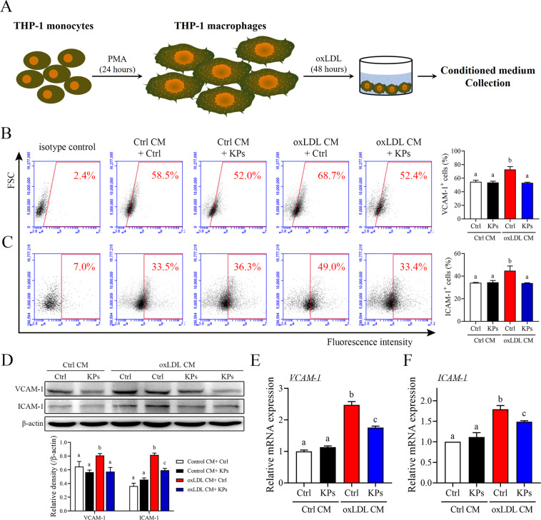
Figure 6.
Effects of kefir peptides on endothelial adhesion molecules. (A) Flow chart of the conditioned medium (oxLDL CM) collection from ox-LDL-stimulated THP-1 macrophages. Conditioned medium from unstimulated THP-1 macrophages were used as a control (Ctrl CM). (B, C) Expression of adhesion molecules, VCAM-1 and ICAM-1, in HUVECs after 6 h incubation in oxLDL CM or Ctrl CM with or without kefir peptides (KPs, 100 μg/ml) were determined by flow cytometry. The histograms on the (B and C) right show the quantification data of three independent HUVECs samples of VCAM-1 or ICAM-1-antibody staining, respectively. (D) Western blot analysis of VCAM-1 and ICAM-1 protein expressions. The histogram on the lower panel shows the quantitative densitometry data of the Western blot analysis determined by ImageJ. Quantitative mRNA expressions of VCAM-1 (E) and ICAM-1 (F) were performed by real-time RT-PCR analysis. Values were normalized to the β-actin gene and are expressed relative to the control (Ctrl) group. The statistical analysis was performed according to Duncan’s multiple-range method. The labels at the top of columns without the same letters indicate significant differences between groups (P < 0.05).

