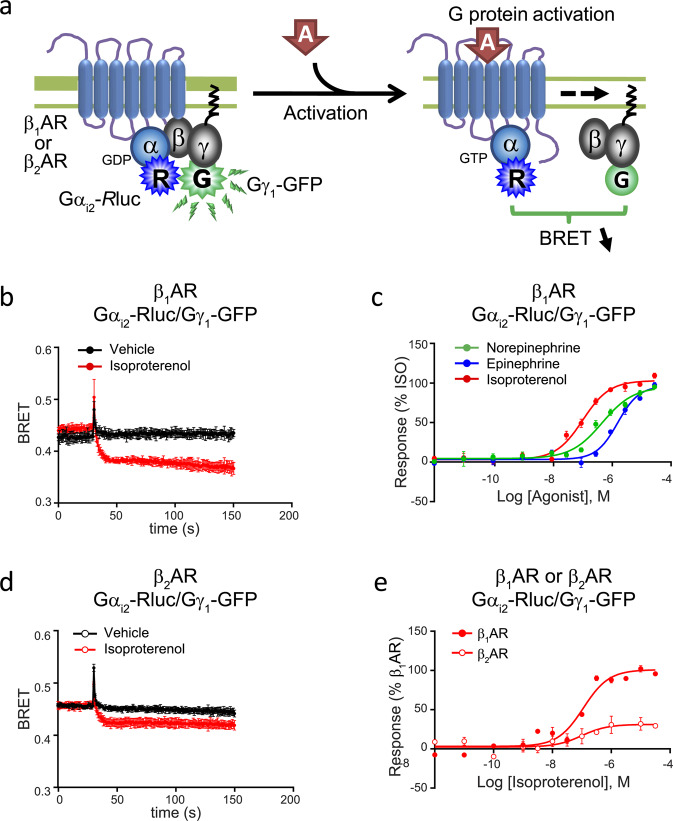
Figure 3.
Gαi2-induced activation by the β1AR and β2AR. (a) Schematic representation of the Gαi2-Rluc/Gγ1-GFP biosensor used to study the Gαi induced β1AR and β2AR signalling. HEK 293 cells were transfected with (b-e) Gαi2-Rluc, Gγ1-GFP and untagged Gβ1, along with (b-c,e) β1AR or (d-e) β2AR. (b,d) Kinetics curves represent time course of Gαi2 activation by (b) β1AR (vehicle and isoproterenol, n=3) or (d) β2AR (vehicle and isoproterenol, n=3) expressed as absolute BRET ratios. (c) Concentration-responses curves for Gαi2 activation following β1AR activation by indicated ligands. (e) Concentration-responses curves for Gαi2 activation following isoproterenol-induced activation of β1AR or β2AR (n=2). Data were normalized to maximal isoproterenol response, which was take as 100%, and are expressed as mean ± SEM values. Detail (c) of the number of experiments, maximal responses, pEC50 values and statistical comparisons of curve parameters for different ligands are provided in Supplementary Tables S1 and S2.

