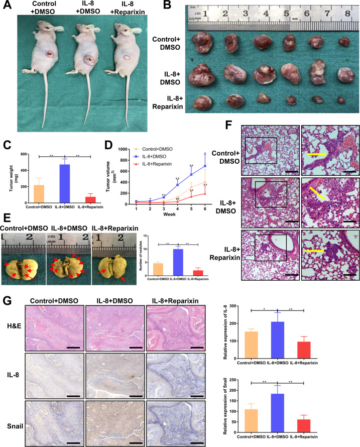
Fig. 4. IL-8 facilitates HNSCC progression in vivo through CXCR1/2.
a, b Representative images of xenograft tumors derived from mice treated with IL-8 (0.5 mg/kg, every other day) + DMSO, IL-8 (0.5 mg/kg, every other day) + Reparixin (15 mg/kg, once a day), or PBS + DMSO; (n = 6). c Tumor weight was measured after excision from mice in three groups. d Tumor volume was examined every week and plotted as tumor growth curves. IL-8 + DMSO group and IL-8 + Reparixin group were compared with the control group. e Representative lung tissues from different groups in the metastasis assay were fixed in Bouin’s fixative diluted 1:5 with neutral-buffered formalin; (n = 6). Arrows indicate metastatic nodules. f H&E staining of lung tissues was performed to examine the pathologic type of the metastatic nodules (magnification 100×, 200×). Scale bar, 100 μm and 50 μm. Arrows indicate metastatic cell nests. g H&E and IHC staining for IL-8, and snail were conducted on xenograft tumors (magnification 100×). Scale bar, 100 μm; *P < 0.05, **P < 0.01.

