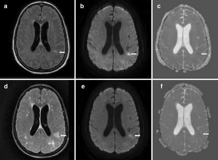
Fig. 1.
MRI brain on hospital day 29 showed FLAIR hyperintensities in the deep hemispheric, periventricular, and juxtacortical white matter (arrow, a), mostly hyperintense on diffusion weighted imaging (DWI) (arrow, b), and some show subtle restricted diffusion on apparent diffusion coefficient (ADC) imaging (arrow, c). Repeat MRI brain on hospital day 58 showed an increased number and distribution of FLAIR hyperintensities in the hemispheric white matter (arrow, d), with persistence of some previous lesions on DWI (arrow, e) and ADC (arrow, f), but no new lesions on these sequences

