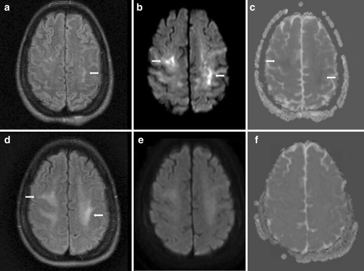
Fig. 2.
MRI brain on hospital day 24 showed small FLAIR hyperintensities in the juxtacortical white matter (arrow, a), more widespread hyperintensities on diffusion weighted imaging (DWI) (arrows, b), with subtle restricted diffusion on apparent diffusion coefficient (ADC) imaging (arrows, c). Repeat MRI brain on hospital day 58 showed poorly defined FLAIR hyperintensities in the juxtacortical white matter (arrow, d), with resolution of the signal abnormalities on DWI (e) and ADC (f) sequences

