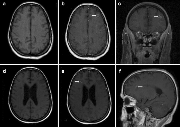
Fig. 3.
MRI brain on hospital day 24 showed a small contrast enhancing lesion (arrows) in the left frontal lobe at the grey-white interface (a T1 axial. b T1 axial post-gadolinium. c T1 coronal post-gadolinium). MRI brain on hospital day 58 showed a small contrast enhancing lesion (arrows) in the right frontal white matter (d T1 axial, e T1 axial post-gadolinium, f T1 sagittal post-gadolinium.)

