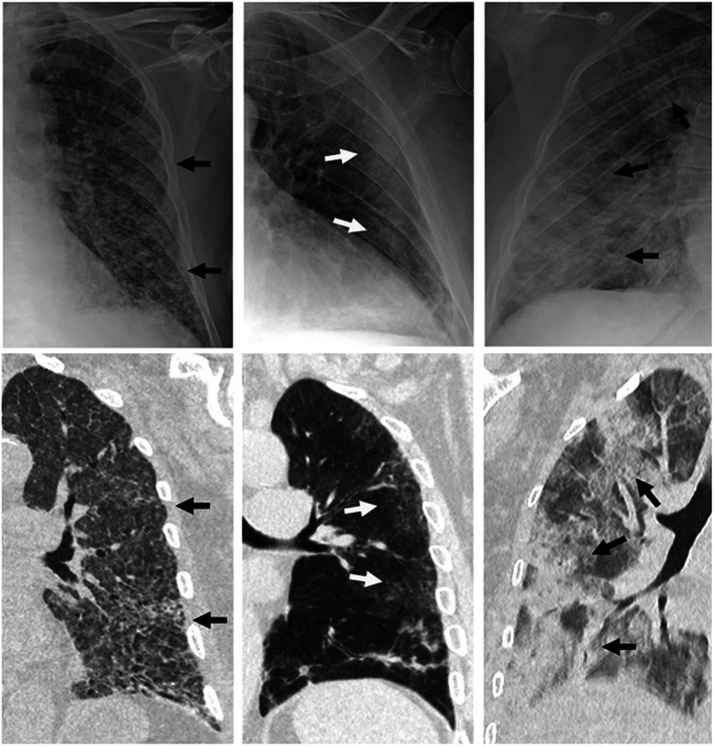
Fig. 1.
The three main alterations on chest radiography (upper line) and the corresponding findings on chest CT (lower line). Left: diffuse reticular alteration (arrows). The corresponding CT shows diffuse increased lung attenuation and interlobular septal thickening (arrows). Middle: peripheral ground-glass opacities (arrows). Right: extensive consolidations (arrows). The corresponding CT shows predominant consolidative alterations (arrows)

