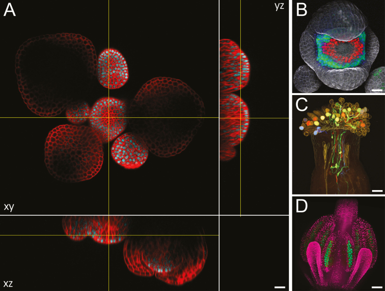
Fig. 1.
Arabidopsis flowers imaged with optical sectioning techniques. (A) Optical xy section and reconstructed xz and yz sections of a live Arabidopsis inflorescence expressing a transcriptional SHOOT MERISTEMLESS reporter (cyan) imaged with a point-scanning confocal microscope; cell walls were stained with propidium iodide (red); these images show the limitation of imaging depth with confocal microscopy. (B) Maximum intensity projection of a live, stage 5 Arabidopsis flower expressing a transcriptional reporter for APETALA3 (AP3; green) and translational reporters for AP3 (green) and SUPERMAN (red); cell walls were stained with propidium iodide (gray); note the differences in expression of the transcriptional and translational AP3 reporters. (C) Maximum intensity projection of an Arabidopsis pistil pollinated with pollen expressing different transcriptional reporters (mTFP1, sGFP, Venus, and mApple) for LAT52, treated with ClearSee for 5 months, and imaged with two-photon excitation microscopy; this image, courtesy of Drs Yoko Mizuta and Daisuke Kurihara, was originally published in Kurihara et al. (2015). (D) Maximum intensity projection of a live Arabidopsis floral bud expressing reporters for the ASY1 (green) and H2B (pink) genes; sepals were removed; image courtesy of Sona Valuchova and Pavlina Mikulkova. Scale bars=50 µm in (A–C), 100 µm in (D).

