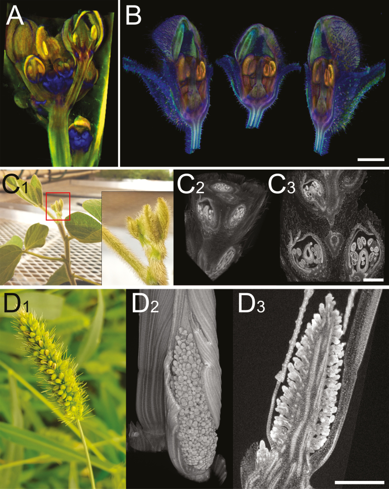
Fig. 2.
Computed tomography images of flowers. (A) Transmission OPT image of an Arabidopsis inflorescence expressing a GUS reporter for LEAFY (blue); image courtesy of Karen Lee. (B) Three views of an Antirrhinum flower imaged with emission OPT and virtual dissecting with a clipping plane to reveal internal structures; image courtesy of Karen Lee. C1–C3. Photograph (C1) and XRM images (C2 and C3) of a young soybean axillary bud containing numerous young florets that will eventually develop into soybean pods; C2 shows a virtual dissection with three clipping planes; C3 shows a computationally reconstructed section. (D1–D3) Photograph (D1), 3D computed reconstruction (D2), and computationally reconstructed section (D3) of young inflorescences from foxtail millet (Setaria viridis). Scale bars=500 µm.

