Fig. 1.
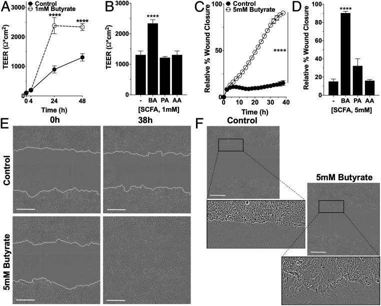
Influence of SCFAs on barrier formation and wound healing. (A) TEER measurements over time in T84 cells on permeable inserts treated with 1 mM butyrate (n = 6; error bars: SEM, ****P < 0.0001 by two-way ANOVA, Fisher’s multiple comparison). (B) TEER measurement at 48 h of T84 cells treated with 1 mM butyrate (BA), propionate (PA), or acetate (AA) (n = 6; error bars: SEM, ****P < 0.0001 by one-way ANOVA, Fisher’s multiple comparison). (C) Scratch wound healing monitored over time by relative wound closure percentage in T84 cells treated with 5 mM butyrate (n = 5; error bars: SEM, ****P < 0.0001 by two-way ANOVA, Fisher’s multiple comparison). (D) Relative wound closure percentage at 38 h of T84 cells treated with 5 mM BA, PA, or AA (n = 5; error bars: SEM, ****P < 0.0001 by 1-way ANOVA, Fisher’s multiple comparison). (E) Live cell images of scratch wound healing (0 h, 38 h) in T84 control cells and cells treated with 5 mM butyrate (gray, cell migration/wound edge). (Scale bar: 300 μm, 10×.) (F) Live cell images of the migratory edge at 6 h in control and T84 cells treated with 5 mM butyrate. (Scale bar: 300 μm, 10×.)

