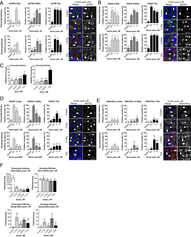
Fig. 1.
The DNA repair response is activated in prophase-arrested oocytes following DNA damage. WT mice (PN10) were exposed to whole-body γ-irradiation at 0.2, 4.5, or 7 Gy. Prophase-arrested oocytes in primordial and growing follicles were analyzed in untreated controls 0.5, 3, and 6 h later for nuclear localization of pATM (A), γH2AX (B and C), RAD51 (D), and DNA-PKcs (E). Representative immunofluorescence images are shown for controls and at 0.5 h post γ-irradiation. DNA was counterstained with DAPI (blue), and oocytes were labeled with c-Kit or MVH (green) and colocalized with markers of the DNA damage response and repair (red; arrowheads indicate oocytes with positive staining, and arrows indicate negative staining) (Scale bars, 20 µm.) (F) Ovaries were also collected 24 h after γ-irradiation, and oocytes were enumerated (n = 4 to 5 mice/group). All data are expressed as mean ± SEM and analyzed by one-way ANOVA followed by Tukey’s post hoc test or Kruskal–Wallis for nonparametric data. Different letters are significantly different; P < 0.05. Localization of each marker was evaluated in oocytes from ∼50 to 200 primordial follicles and ∼25 to 100 growing follicles per treatment and time point; n = 3–8 animals/group.

