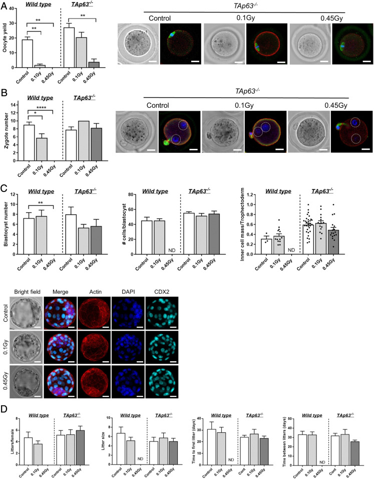
Fig. 3.
DNA repair is sufficient to restore functional fertility. (A) Number of ovulated oocytes harvested from γ-irradiated wild-type and TAp63−/− mice following exogenous hormonal stimulation. Representative images of oocytes obtained from TAp63−/− females stained with f-actin (red) to mark the oolema, αβ-tubulin (green) to label the spindle, and DAPI to label the DNA on the metaphase plate. (B) Number of zygotes obtained from γ-irradiated WT and TAp63−/− mice at E0.5 after mating with untreated males. Representative images of zygotes obtained from TAp63−/− females stained with f-actin (red) to mark the zygote boundary and with DAPI to label the DNA in pronuclei (circled by dotted white lines) to confirm fertilization. (Scale bar, 20 μm.) (C) Number of blastocysts obtained from γ-irradiated WT and TAp63−/− mice at E3.5 after mating with untreated males. The ratio of inner cell mass to trophectoderm (TE) is shown. Representative images of blastocysts obtained from TAp63−/− females stained with f-actin (red) to mark the cell boundaries and with DAPI to label the DNA and CDX2 (aqua) to label TE. (Scale bar, 20 μm.) Fertility of mice (D) from continuous mating trial. All data are expressed as mean ± SEM and analyzed by one-way ANOVA followed by Tukey’s post hoc test or Kruskal–Wallis for nonparametric data or t tests for pairwise comparisons. Different letters are significantly different; *P < 0.05, **P < 0.005, ****P < 0.0001.

