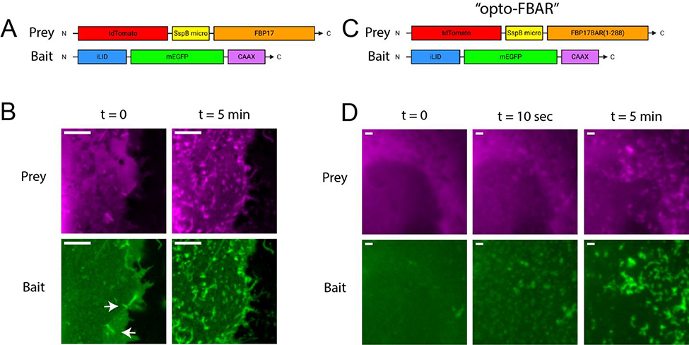
Figure 2:
The construction and optimization of opto-FBAR. A) Box drawing of the initial design of the opto-FBAR system using full length FBP17. B) Zoomed images of COS7 cells co-expressing tdTom-SspBmicro-FBP17 and iLID-EGFP-CAAX shown in A. In the dark (t = 0), cells show some FBP17-positive membrane tubules (arrows). Blue light stimulation (t = 5 min) drastically increased the amount of membrane tubules that is positive in both channels. Full field images available in Supplementary Figure S2. Scale bar = 5 μm. C) Box drawing of final opto-FBAR system using the FBAR domain, i.e. truncated FBP17 (a.a. 1–288). D) Zoomed images of COS7 cells expressing the final opto-FBAR system shown in C. tdTom-SspBmicro-FBAR is completely cytosolic in the dark. Blue light rapidly recruits the prey to the membrane and results in the formation of membrane tubules positive in both channels. Full field images available in Supplementary Figure S4. Scale bar = 1 μm.

