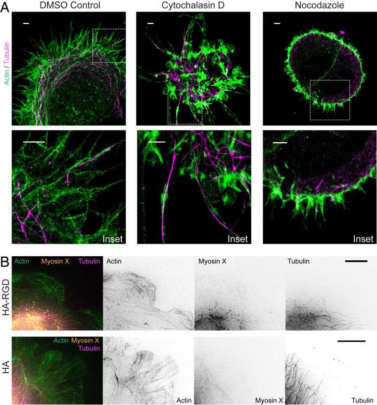
Fig. 4.
Organization of microtubules and actin within McTNs. (A) STORM imaging of U-251 MG cells treated with DMSO as a control shows actin filaments extending around the cell periphery with microtubules interspersed and extending to the ends of McTN tips. After cytochalasin D treatment, most protrusions contain microtubules at the core. Cells treated with nocodazole adhere poorly to HA, remain rounded, and express only short, actin-positive protrusions at the periphery. Dashed line boxes represent Inset area. Insets show protrusions at cell periphery. (Scale bars: 2 µm.) (B) U-251 MG cells on bare HA do not express myosin X at the cell periphery. Cells on HA-RGD have some myosin X at the leading edge compared to cells on HA. (Scale bars: 5 µm.)

