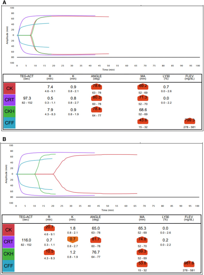
Fig. 2.
a Thromboelastography (TEG) performed at diagnosis of acute ischaemic limb. CK angle 78.6° (63°–78°) and CRT angle 80.3° (60°–78°) are increased, CK MA 69.2 mm (52–69 mm) and CRT MA 71.3 mm (52–70 mm) are also increased, CFF MA 43.8 mm (15–32 mm) is markedly increased secondary to hyperfibrinogenaemia, reflected by high functional fibrinogen (FLEV) 799.3 mg/dl. b Thromboelastography (TEG) on post-operative day 1 performed on citrated blood sample with IV heparin infusion. CK R is markedly prolonged 20.7 min (4.6–9.1 min) due to the heparin effect, while CKH R is borderline prolonged 10.5 min (4.3–8.3 min). CKH MA is still raised at 69.3 mm (52–69 mm), suggestive of a persistent hypercoagulable state in the absence of heparin, while CK MA is normal, suggestive that clot formation is normalised in presence of heparin. CFF MA 53.8 mm (15–32 mm) is further increased secondary to hyperfibrinogenaemia, reflected by higher functional fibrinogen (FLEV) 981.8 mg/dl which may be reflective of the postoperative infective systemic inflammatory milieu

