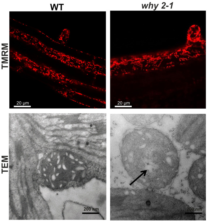
FIGURE 3.

Mitochondria morphology in plant tissues. Upper panel: confocal images of roots of WT and why 2‐1 plants stained with TMRM. Lower panel: transmission electron microscope images of mitochondria from leaf section from 3‐week‐old WT and mutant plants. Arrow indicates translucent area within mitochondria matrix
