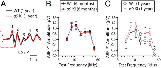
Fig. 3.
Suprathreshold response amplitude for ABR peak 1 in young and aged WT and α9KI mice. (A) Representative ABR waveforms (32 kHz, 80 dB SPL) from WT (black) and α9KI (red) mice at 1 y of age showed a large reduction in wave 1 amplitudes. Dashed lines mark peak 1 amplitude from aged WT (black) and α9KI (red) mice. (B) ABR peak 1 amplitudes at 80 dB SPL in WT (n = 10) and α9KI (n = 10) mice at 6 mo of age at different test frequencies. (C) ABR peak 1 amplitudes at 80 dB SPL in WT (n = 6) and α9KI (n = 6) mice at 1 y of age. Aged WT ears displayed a significant reduction in peak 1 amplitudes at high frequencies. Group means ± SEM. Asterisks represent the statistical significance (Mann–Whitney U test, *P < 0.05).

