Fig. 1.
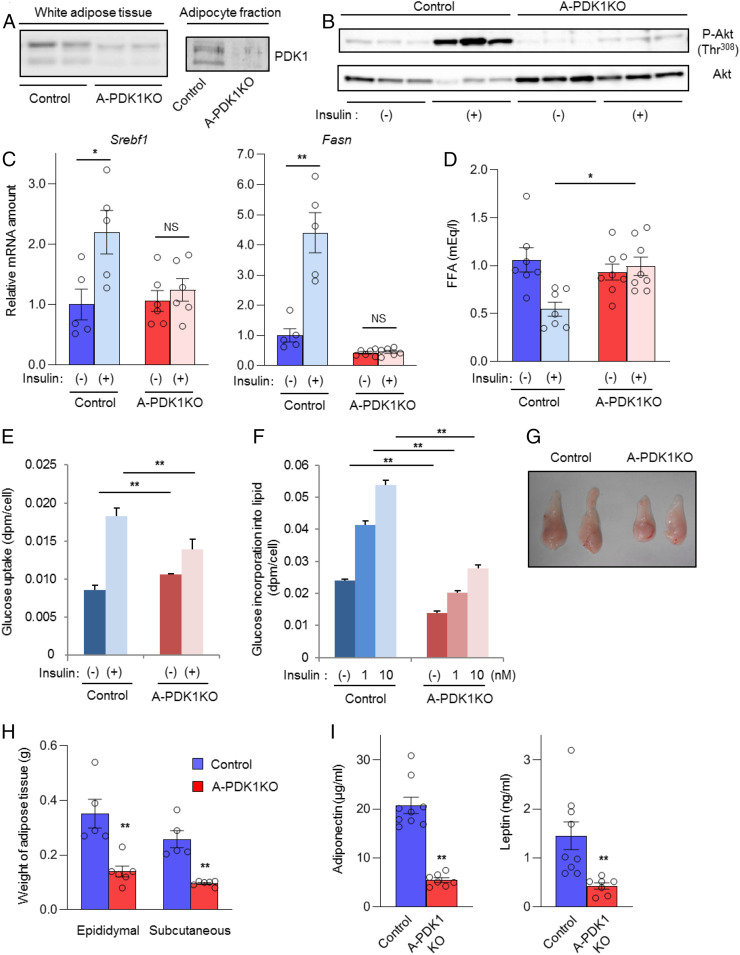
Impaired insulin action in adipocytes of A-PDK1KO mice. (A) Immunoblot analysis of PDK1 in epididymal adipose tissue and in the adipocyte fraction of this tissue from control (PDK1flox/flox) and A-PDK1KO mice. (B) Immunoblot analysis of total and Thr308-phosphorylated (P-) forms of Akt in epididymal adipose tissue isolated from control or A-PDK1KO mice (n = 3) at 10 min after i.p. injection of insulin (5 U/kg) or vehicle. (C) RT and real-time PCR analysis of Srebf1 and Fasn expression in epididymal adipose tissue isolated from control or A-PDK1KO mice (n = 5 or 6) at 6 h after i.p. injection of insulin (1 U/kg) or vehicle. (D) Plasma FFA concentration in control or A-PDK1KO mice (n = 7 or 8) measured 1 h after i.p. injection of insulin (1 U/kg) or vehicle. (E) Basal and insulin (1 nM)-stimulated glucose uptake in isolated adipocytes from control or A-PDK1KO mice (n = 3). (F) Basal and insulin-stimulated lipogenesis in isolated adipocytes from control or A-PDK1KO mice (n = 3). (G) Representative images of epididymal adipose tissue and (H) weight of epididymal or subcutaneous (s.c.) adipose tissue from control or A-PDK1KO mice (n = 5 or 6). (I) Plasma adiponectin and leptin concentrations in control or A-PDK1KO mice (n = 7 to 9). All quantitative data are means ± SEM; *P < 0.05, **P < 0.01 for the indicated comparisons or versus corresponding control value (Student’s t test); NS, not significant.

