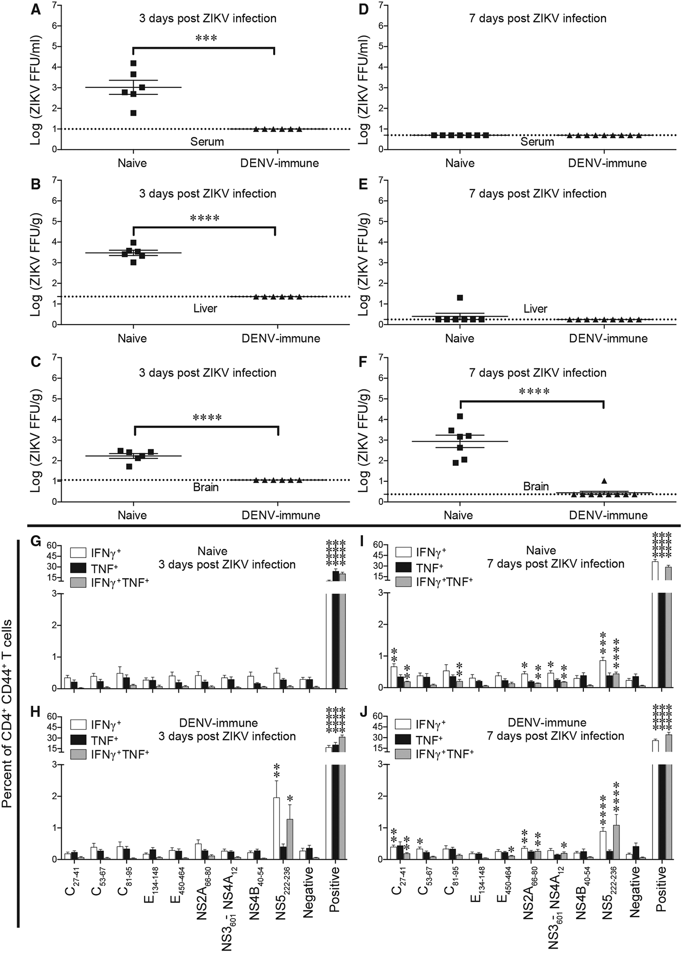
Figure 3. Reduction in Viral Load in DENV-Immune Mice after ZIKV Infection.

Ifnar1−/−HLA-DRB1*0101 mice were infected intraperitoneally with 2 × 10³ FFU of DENV2 S221 for 4 weeks. Naive mice and DENV2-immune mice were infected retro-orbitally with 1 × 10⁴ FFU of ZIKV strain SD001 for 3 (A–C and G–H) or 7 (D–F and I–J) days.
(A–F) Mouse blood (A and D), liver (B and E), and brain (C and F) were harvested, and levels of infectious ZIKV were determined using the FFA. Each point represents an individual mouse.
(G–J) Splenocytes were isolated from mice at 3 dpi (G and H) or 7 dpi (I and J) and stimulated in vitro for 6 h with the indicated ZIKV-specific and DENV2/ZIKV-cross-reactive peptides, and the frequencies of CD4+ T cells producing IFNγ, IFNγ plus TNF, or TNF were detected using the ICS assay. Negative and positive refer to cells incubated alone or with PMA and ionomycin, respectively. Data represent the mean ± SEM of two independent experiments (n = 3–5 mice/experiment).
(A–F) ***p < 0.001, ****p < 0.0001 by two-tailed Mann-Whitney test.
(G–J) *p < 0.05, **p < 0.01, ***p < 0.001, ****p < 0.0001 by one-tailed Mann-Whitney test with a B-H adjusted p value (FDR) < 10%.
