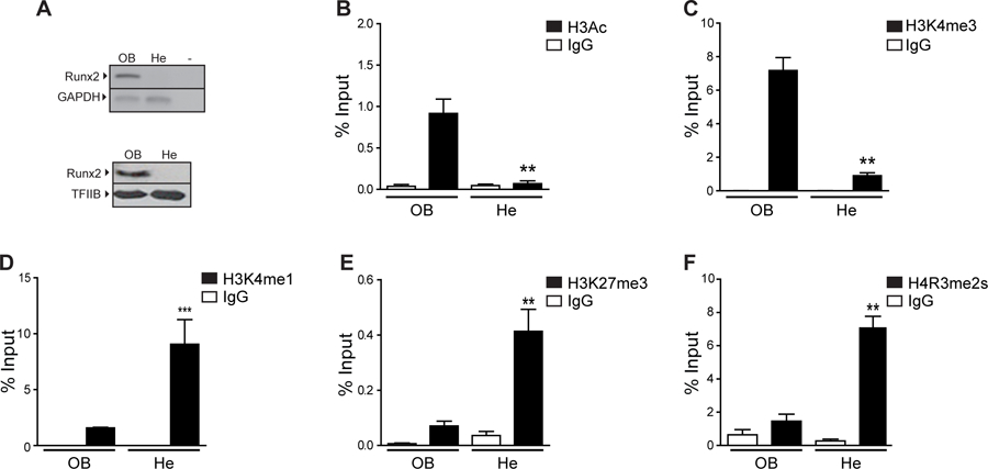
Figure 1. Histone modifications at the Runx2 P1 promoter in osteoblastic and non-osteoblastic cells.

(A) Runx2/p57 expression at mRNA (up) and protein (down) levels was analyzed by RT-PCR and western blot, respectively in samples obtained from osteosarcoma (ROS17/2.8; OB) or hepatoma (H-4-II-E; He) rat cells. Gapdh mRNA is shown as mRNA expression control. Detection of TFIIB was used to control for equal protein loading. (B-F) Enrichment of histone modifications at the Runx2 P1 promoter in osteosarcoma or hepatoma rat cells. Chromatin immunoprecipitation (ChIP) assays were performed using specific antibodies against the histone modifications (B) H3Ac, (C) H3K4me3, (D) H3K4me1, (E) H3K27me3 and (F) H4R3me2s. ChIP values are expressed as % Input ± SEM. Normal IgG was used as specificity control. Statistical analyses were performed respect to ChIP-values obtained from ROS17/2.8 cells. ***p<0.001, **p<0.01.
