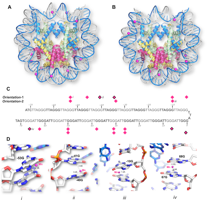
Figure 1.
The two-orientation telomeric NCP crystal structure solved by a combination of molecular replacement, and single-wavelength anomalous maps at the phosphorous, manganese and cobalt absorption edge. Mn2+ coordination to the N7 guanine atoms in (A) Telo-NCP-1 (Orientation-1) and (B) Telo-NCP-2 (Orientation-2). (C) Coordination pattern of the Mn2+ atoms in Telo-NCP-1 and Telo-NCP-2. The Mn2+ shows two modes of coordination: Mn2+ bound to a single N7 guanine (shown as magenta diamonds), and Mn2+ interacting with N7 of two adjacent guanine bases (marked as magenta diamonds with black borders). (D) Examples of Mn2+ interaction with DNA are shown in panels iii, iii and iv. Panels i and iii show Mn2+ coordination to a single N7 guanine in Telo-NCP-1 and Telo-NCP-2 respectively. Panels ii and iv display Mn2+ interaction with N7 of two adjacent guanine bases in the Telo-NCP-1 and Telo-NCP-2 respectively. Henceforth, for illustration, the most frequent naming of DNA and proteins chains is adopted. The two DNA strands are named as ‘I’ and ‘J’, with nucleotides on each strand numbered from −72 bp to 72 bp. The nucleotides are labelled with a numeral identifier followed by strand identifier, e.g. ‘−2I’ corresponding to nucleotide number ‘−2’ from the strand I and histones are labelled A and E (H3) (blue); B and F (H4) (green); C and G (H2A) (yellow); and D and H (H2B) (red).

