Figure 6.
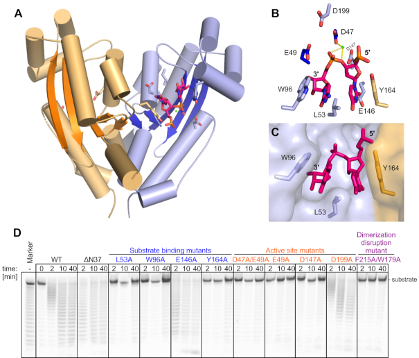
Crystal structure of REXO2 (28–226) and verification of residues involved in catalytic activity. (A) The two protomers are in orange and blue with β-strands in darker shades of the same color. The RNA molecule, active site residues, and residues that are involved in RNA binding are shown as sticks. The Ca2+ ion is shown as a green sphere. (B) Magnification of the active site and residues that are involved in RNA binding. Tyr164 from the other protomer of the dimer is shown in orange. (C) Aromatic clamp showing the first two nucleotides from the 3′ end positioned between W96 from one molecule and Y164 from the other molecule. The protein is shown in surface representation, and two nucleotides of the RNA and selected protein residues are shown as sticks. (D) Poly(U)25 digestion by different mutants of REXO2.

