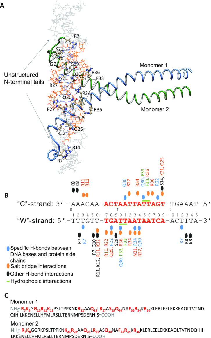
Figure 6.

Model structure and interactions network of the Yap8–DNA complex. (A) Model structure of Yap8 protein homodimer in complex with the 25 bp long DNA segment containing Yap8 response element (Y8RE), in orange. Each Yap8 monomer, shown with labeled major DNA-interacting residues, includes an unstructured N-terminal region (residues 7–16) and basic leucine zipper domain (residues 17–89). (B) Schematic overview of the protein–DNA interactions. The DNA sequence, used in the model, is numbered 1–25 with the ‘Watson’ (‘W’) strand representing the 5′-3′ direction and the ‘Crick’ (‘C’) strand – the 3′-5′ direction. Only the interactions that occur at least 25% of the time of the 0.5 μs MD simulation are depicted. (C) Amino-acid sequences, residues 7–89, of Yap8 monomers included in the model. In bold-red are the residues that show the stable interactions with DNA.
