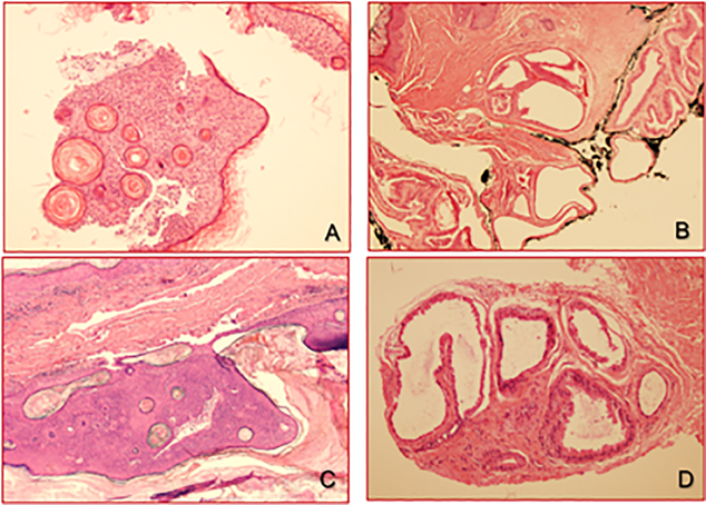
Figure 1.
Right auditory canal biopsy showing an irritated seborrheic keratosis in 2003 (A); at the same location, an apocrine hidrocystoma was excised in 2007, extending to the inked margin (B); after 7 years, recurrent pigmented seborrheic keratosis (C) and adjacent apocrine hidrocystoma (D) at the same right auditory canal.

