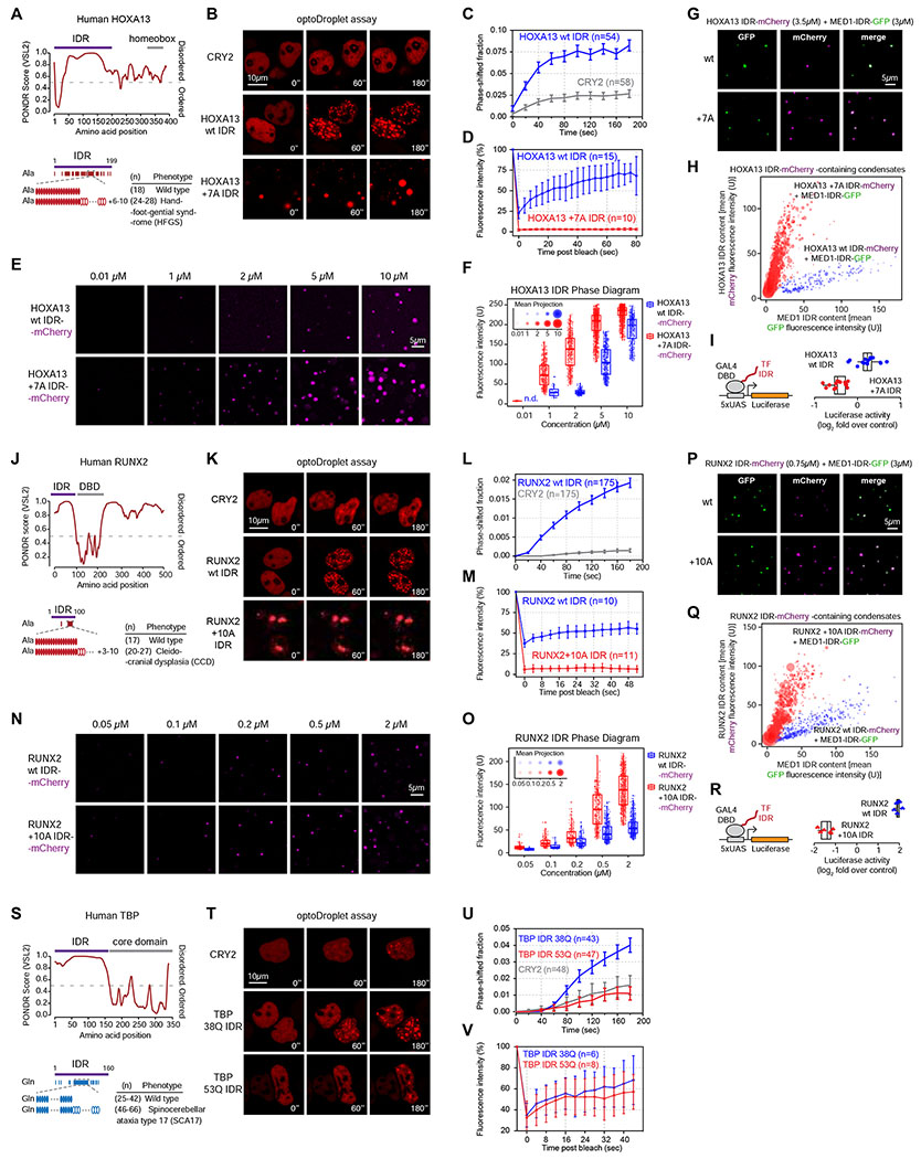
Figure 6. Disease-associated repeat expansions alter the phase separation capacity of other TF IDRs.
(A, J, S) Graphs plotting intrinsic disorder for HOXA13, RUNX2 and TBP. The IDRs cloned for subsequent experiments are highlighted with a purple bar.
(B, K, T) Representative images of HEK-293T nuclei expressing the indicated TF IDR-mCherry-CRY2 fusion proteins. Cells were stimulated with 488nm laser every 20s for 3 minutes.
(C, L, U) Quantification of the fraction of the nuclear area occupied by droplets of the indicated TF IDR-mCherry-CRY2 fusion proteins in HEK-293T nuclei over time. Data displayed as mean+/−SEM.
(D, M, V) Fluorescence intensity of droplets of the indicated TF IDR-mCherry-CRY2 fusion proteins before, during and after photobleaching. For the HOXA13 +7A IDR and the RUNX2 +10A IDR the spontaneously formed droplets were bleached, for all other fusion proteins the light-induced droplets were bleached. Data displayed as mean+/−SD.
(E, N) Representative images of droplet formation by purified TF IDR-mCherry fusion proteins in droplet formation buffer.
(F, O) Phase diagram of TF IDR-mCherry fusion proteins. Every dot represents a detected droplet. The inset depicts the projected average size of the droplets as mean+/− SD (middle circle: mean, inner and outer circle: SD). n.d.: not detected
(G, P) Representative images of droplet formation by purified MED1-IDR-GFP and TF IDR-mCherry fusion proteins in droplet formation buffer with 10% PEG-8000.
(H, Q) Quantification of GFP and mCherry fluorescence intensity in TF IDR-mCherry containing droplets in the indicated MED1 IDR-GFP mixing experiments. Each dot represents one droplet, and the size of the dot is proportional to the size of the droplet.
(I, R) (left): GAL4 activation assay schematic. The luciferase reporter plasmid, and the expression vector for the GAL4 DBD-TF IDR fusion proteins were transfected into HEK-293T cells. (right): Luciferase reporter activity of the indicated TF IDRs fused to GAL4-DBD. p <10−3 for both wt/mutant comparisons (Student’s t-test).
See also Figure S6.

