
Figure 1.
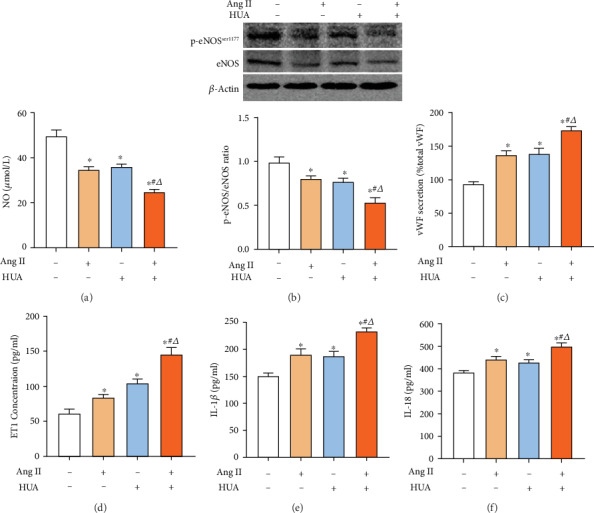
UA and Ang II induce endothelial injury. (a) The NO level of the supernatant was assayed by NO reduction. HUVECs were treated with 600 μmol/L UA (HUA), 10−7 mol/L Ang II (Ang II), or 600 μmol/L UA together with 10−7 mol/L Ang II (HUA+Ang II) to reduce NO production after 24 h of stimulation; ∗p < 0.05 compared to the normal control. The NO level in the HUA+Ang II group was significantly lower than that in the HUA group or the Ang II group; #p < 0.05 compared to the Ang II group; △p < 0.05 compared to the HUA group. (b) Western blotting was used to detect eNOS expression levels in HUVECs. The HUA, Ang II, and HUA+Ang II groups showed decreased phosphorated eNOS-ser1177 protein expression levels in HUVECs after 24 h of stimulation; ∗p < 0.05 compared to the normal control group. The phosphorated eNOS-ser1177 protein expression level in the HUA+Ang II group was significantly lower than that in the HUA group or the Ang II group; #p < 0.05 compared to the Ang II group; △p < 0.05 compared to the HUA group. (c–f) The vWF, ET1, IL-1β, and IL-18 concentration of the supernatant was assayed. HUA and Ang II increased vWF, ET1, IL-1β, and IL-18 production after 24 h of stimulation; ∗p < 0.05 compared to the normal control. The vWF, ET1, IL-1β, and IL-18 in the HUA+Ang II group were remarkably higher than those in the HUA group or the Ang II group; #p < 0.05 compared to the Ang II group; △p < 0.05 compared to the HUA group.