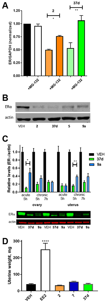
Figure 4.

B-SERD vs SERD comparisons. (A). ERα degradation after 24 h treatment ofMCF7:WS8 cells with 10 nM 37d or 2 measured by western blot and inhibited by proteasomal inhibitor MG-132 (1 μM) normalized to vehicle (1.0). (B) ERα western blots after 24 h treatment of MCF7:WS8 cells with 100 nM 2, 37d, 5, and 9a measured by the western blot. (C) ERα degradation after oral dosing of female mice with vehicle, 37d or 9a, measued by western blot analysis of tissues, with representative immunoblots shown from individual mouse uterus. (D) Uterine weight from juvenile female rats dosed with 2, 7, and 37d, compared to EE2 as a positive control. Cell culture data shown as mean ± SEM from three biological and analytical replicates. Statistical analysis by one-way ANOVA with multiple comparisons (p* < 0.01; *** > 0.001; **** < 0.0001).
