Figure 1.
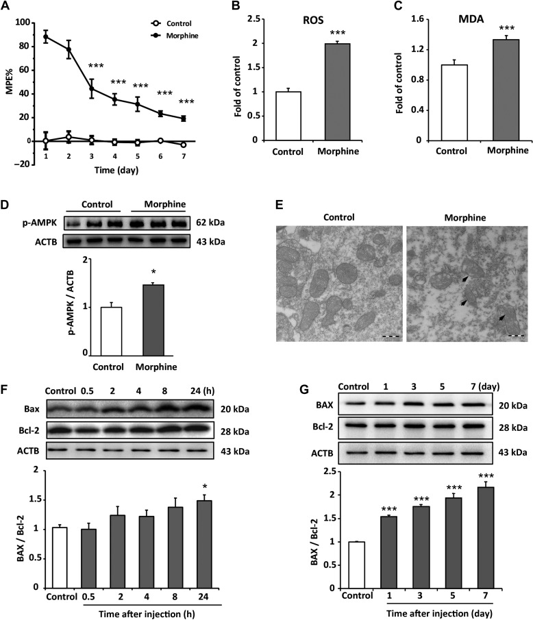
Chronic intrathecal administration of morphine induces excessive generation of ROS and causes accumulation of damaged mitochondria in spinal cord. (A) Tail-flick method was performed to evaluate morphine tolerance. Data are shown as percentage of MPE. Chronic administration reduced morphine’s MPE from Day 3 to Day 7. The saline-treated group served as control. The MPE from Day 3 to Day 7 were 44.5%, 35.5%, 31.4%, 23.4%, and 19.3%, respectively. One-way ANOVA followed by Tukey’s multiple comparisons test. n = 8, ***P < 0.001 vs. MPE of Day 1. (B and C) The levels of ROS and MDA on Day 7 from spinal cord tissue were assessed by DCFH-DA staining and MDA detection kit. The ROS level and MDA level increased by 99.1% and 32.9%, respectively, compared with control group. Student’s t-test. n = 6, ***P < 0.001 vs. control group. (D) Increased phosphorylation of AMPK (Thr172) was detected in morphine-treated group compared with control group. Student’s t-test. n = 4, *P < 0.05 vs. control group. (E) Sections of spinal cord from mice chronically administrated with morphine were fixed and subjected to EM examination. Morphine induced a significant damage and accumulation of abnormal mitochondria compared with control group. Scale bar, 500 nm. (F and G) Morphine increased the ratio of Bax/Bcl-2 after 24 h of administration; in long-term treatment, morphine significantly increased the ratio of Bax/Bcl-2 from Day 1 to Day 7. n = 4, *P < 0.05, ***P < 0.001 vs. control group.

