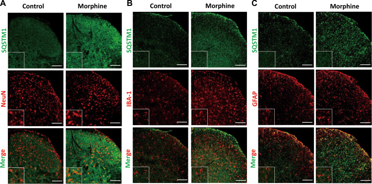
Figure 3.
The distribution and cellular location of SQSTM1/p62 after seven consecutive days of morphine intrathecal injection in the spinal cord dorsal horn. Sections of spinal cord from mice chronically administrated with morphine were fixed and subjected to double immunofluorescence stain. Confocal microscopy was performed to determine the co-localization of SQSTM1/p62 (green) and neuron (NeuN, red), microglia (IBA-1, red), or astrocyte (GFAP, red). Spinal samples were collected after the last administration of morphine. NeuN, neuronal nuclear protein; IBA-1, ionized calcium-binding adapter molecule 1; GFAP, glial fibrillary acidic protein. Scale bar, 100 μm.

