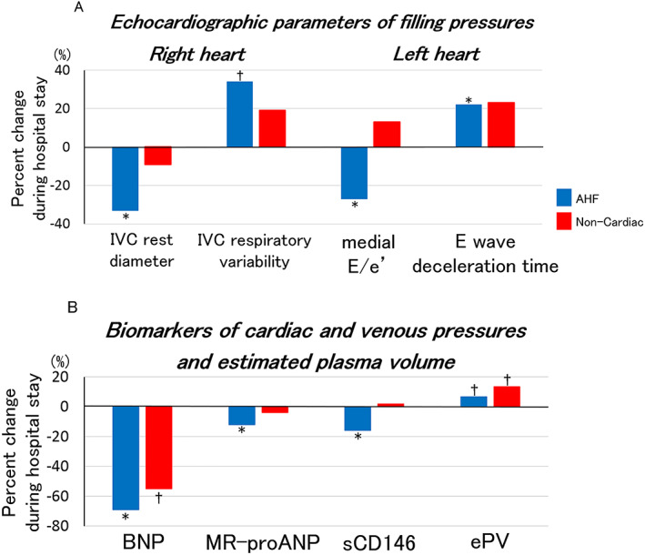
Figure 1.

Percent changes in echocardiographic parameters and plasma biomarkers of cardiac and venous pressures and estimated plasma volume during hospital stay for AHF. Figure 1(A) shows median values of percent changes in echocardiographic parameters of right and left cardiac filling pressures (IVC rest diameter, IVC respiratory variability, medial E/e', and E wave deceleration time) during hospital stay in AHF. Figure 1(B) shows median values of percent changes in plasma biomarkers of cardiac and venous pressures (BNP, MR‐proANP, and sCD146) and estimated plasma volume during hospital stay in AHF. *P < 0.01, †P < 0.05. AHF, acute heart failure; BNP, B‐type natriuretic peptide; ePV, estimated plasma volume; IVC, inferior vena cava; MR‐proANP, mid‐regional pro‐atrial natriuretic peptide; sCD146, soluble cluster of differentiation 146.
