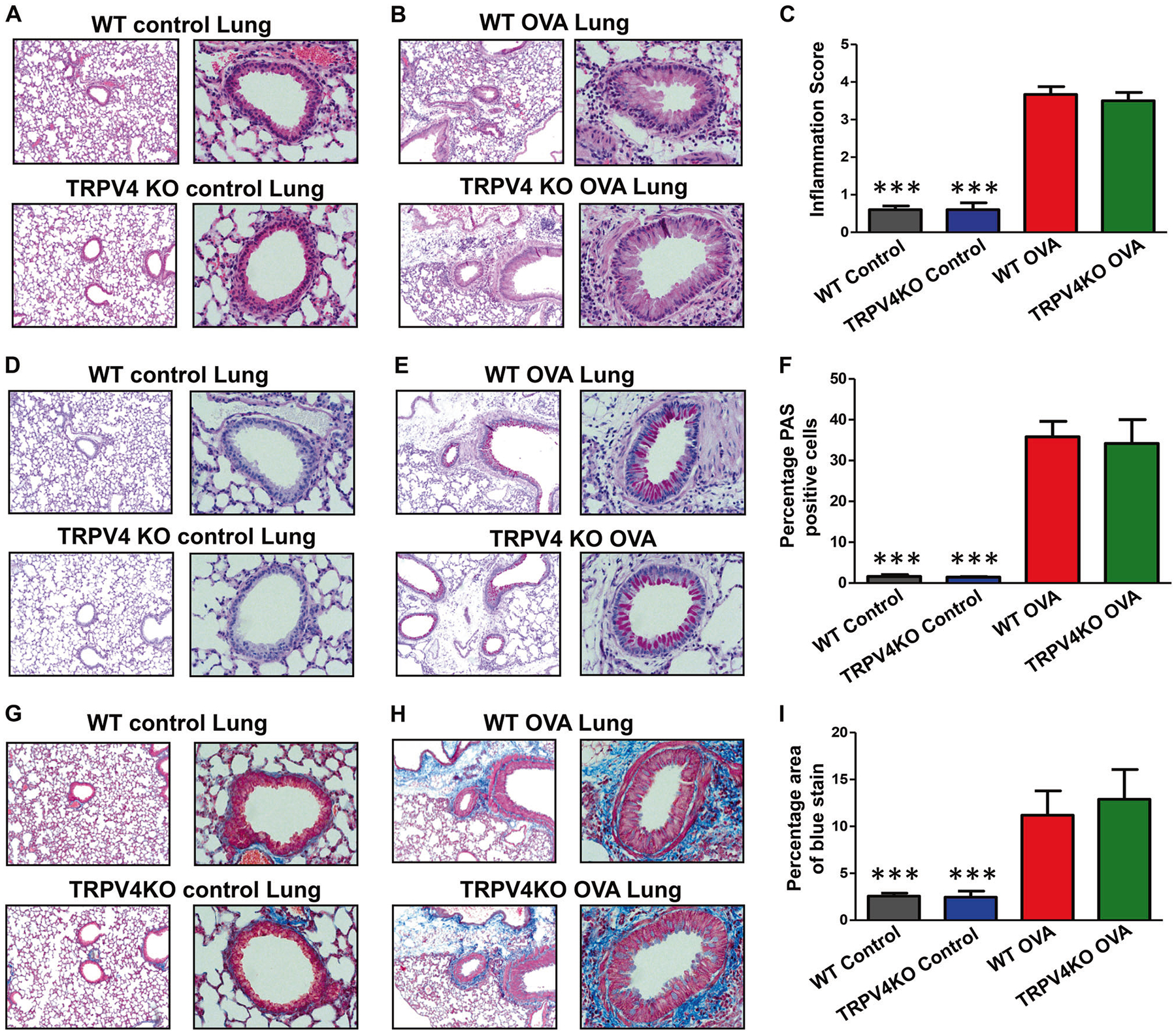
Fig. 5.

Histopathological analysis of airway inflammation. a-i WT and TRPV4 KO mice were OVA-sensitized and challenged with OVA. Experimental mice and untreated control WT and TRPV4 KO mice were euthanized 48 h after last OVA challenge and lung tissues were collected, fixed in buffered formalin, paraffin-embedded, and thin sections were made. Sections were stained with H&E (a, b), periodic acid schiff (d, e), or Masson trichrome (g, h). c, f, i Quantified data (mean ± SEM) from analysis of thin sections prepared from three individual OVA-sensitized and challenged mice stained with H&E (c), periodic acid schiff (f), or Masson trichrome (i). Images are representative of three independent experiments (b, e and h). Similarly, lung sections from untreated WT and TRPV4 KO control mice were also prepared. Quantified data (mean ± SEM) from analysis of thin sections prepared from five individual WT and TRPV4 KO control mice stained with H&E (c), periodic acid schiff (f), or Masson trichrome (i). Data are mean ± SEM of five WT and TRPV4 KO control mice (c, f, i). Image is representative of five individual mice. a, d, g The percentage area of blue color in Masson-trichrome stain for deposition of collagen/lung fibrosis was estimated using ImageJ software
