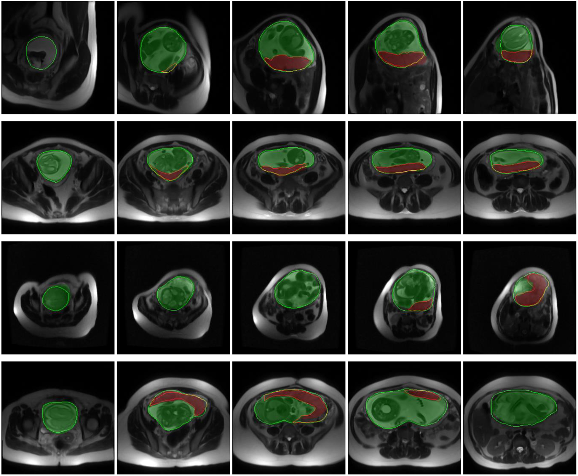
Figure 4.

Qualitative results of the uterus and placenta segmentation in 2D. Each row shows the results for one patient on five sample axial slices from inferior (left) to superior (right). The semi-transparent green (bright) and red (dark) regions show the algorithm segmentation results for uterine cavity and placenta, respectively. The solid green (bright) and red (dark) contours show the manual segmentation provided by the expert observer for uterine cavity and placenta, respectively.
