
Figure 10: Inducible fibroblast-specific Smad3 loss reduces myocardial collagen content.
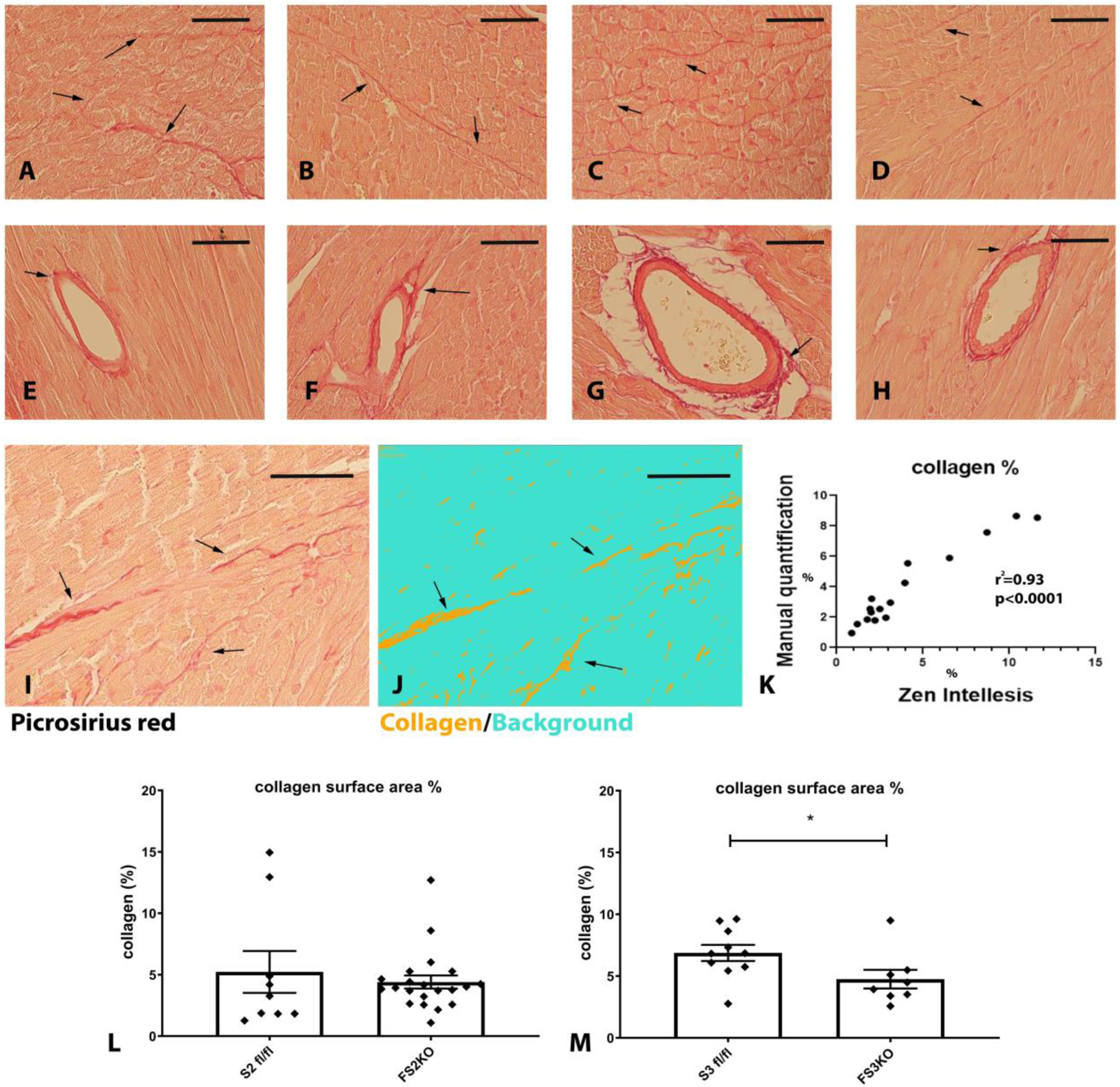
Myocardial sections from Smad2 fl/fl (A, E), FS2KO (B, F), Smad3 fl/fl (C, G) and FS3KO (D, H) mice were stained with picrosirius red to label interstitial (A–D) and perivascular (E–H) collagen fibers (arrows). Mice were studied 8 weeks after tamoxifen injection, FS2KO and FS3KO mice appeared to have preserved myocardial structure. Quantitative analysis of collagen content was performed using a machine learning-based system for objective unbiased analysis. I–K: The artificial intelligence-guided model was tested using 16 different field from 4 mice and showed excellent correlation (r2=0.93, p<0.0001, n=16) with manual measurements. L: FS2KO mice and Smad2 fl/fl controls had comparable myocardial collagen content (p=NS, n=9–21/group, Mann-Whitney test). M: When compared with Smad3 fl/fl animals, FS3KO mice had a modest, but significant reduction in myocardial collagen content (*p<0.05, n=8–10/group, Mann-Whitney test). Scalebar= 50μm.