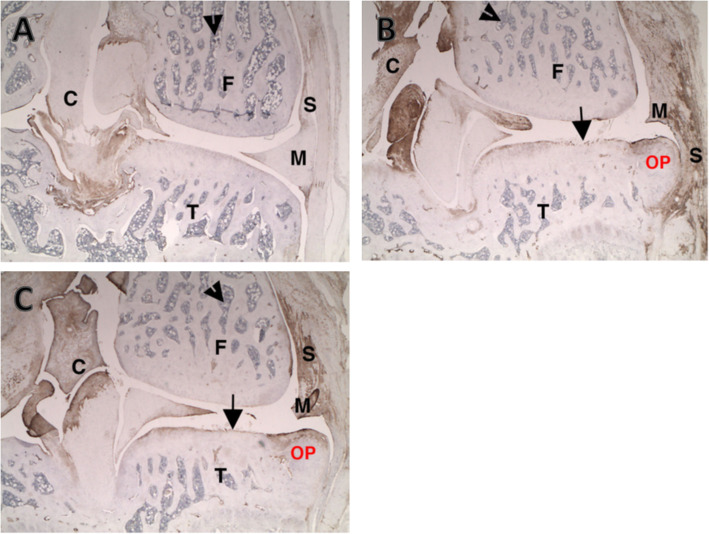
Fig. 6.
TGFβ staining on the medial tibial surface. a Uninjected left knee. Arrowhead identifies bone marrow. Note normal immunopositivity in connective tissue of synovium (S), collateral ligament joint capsule, and cruciates (small C). b PBS injected right knee. Note increased connective tissue staining in healing medial synovium (S) damaged meniscus (M), cruciates (small C), and osteophyte (OP). The damaged tibial cartilage has increased staining of the tangential layer in areas where it is intact. c IA-BioHA (0.5 mg/rat) injected at Days 7, 14, and 21 in the right knee. Note increased connective tissue staining in healing medial synovium (S) damaged meniscus (M), cruciates (small C), and osteophyte (OP). The damaged tibial cartilage has increased staining of the tangential layer in areas where it is intact. F- femur; M – meniscus; T – tibia. Images were acquired at 25x magnification

