
Figure 1.
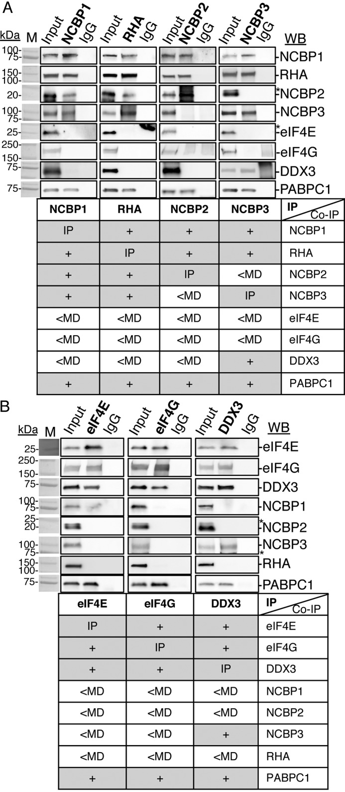
NCBP1-RHA and eIF4E-eIF4G are components of mutually exclusive RNPs. Reciprocal co-IP of selected proteins from HEK293 lysates with specific antisera are shown (bold type). Bound proteins were eluted and analyzed by WB with the antisera indicated on the right. Isotype-specific IgG served as the negative control, and input cell lysate served as the positive control. The results are summarized in the tables below each WB. A, IP of NCBP1, RHA, NCBP2, or NCBP3. B, IP of eIF4E, eIF4G, or DDX3. The antiserum detected the specific proteins on the immunoblots, as shown relative to the prestained molecular mass markers (lanes M). The same image of the molecular mass markers was used for each panel. +, positive co-IP. *, nonspecific background. The WBs were subjected to ImageJ densitometry quantification (Table S1).