
Figure 2.
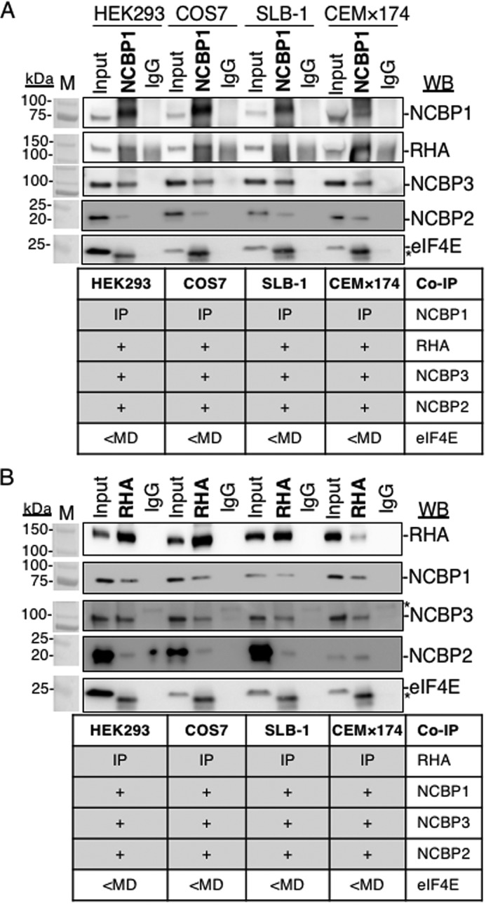
NCBP1-RHA complexes exist in several cell types. IP of NCBP1 (A), RHA (B), or isotype IgG from lysates of HEK293, COS7, SLB-1, or CEM×174 cells depleted of nuclei. Immune complexes (bold type) were washed, collected in SDS sample buffer, and analyzed by WB with the antisera indicated on the right. In each case, the input cell lysate served as the positive control, and the isotype-specific IgG served as the negative control for background immunoreactivity. The results are summarized in the table below each WB. The antiserum detected the specific proteins on the immunoblots, as shown relative to the prestained molecular mass markers (M). The same image of the molecular mass markers was used for each panel. +, positive co-IP. *, nonspecific background. The WBs were subjected to ImageJ densitometry quantification (Table S1).