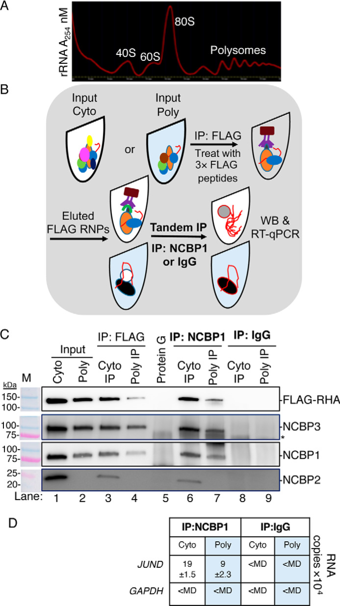
Figure 3.

NCBP1-NCBP3-RHA are components of the same RNP loaded to JUND polysomes. Tandem IPs were employed to isolate components of the same RNP from HEK293 cells transfected with pFLAG-RHA. Input cyto lysates were subjected to sucrose gradient centrifugation, and polysomes were collected. A, A254 spectrometry (red line) of sucrose gradient. B, outline of tandem IP. The polysome fractions were combined, precipitated, and resuspended in low-salt buffer (Input poly). Aliquots of the cyto (white tube) and polysome samples (blue tube) were incubated with FLAG antiserum conjugated to protein G beads. The beads were washed and incubated with 3× FLAG peptides to elute RNPs. A fraction of the eluate was reserved for WB, and the remaining eluate was incubated with NCBP1 antiserum conjugated to protein G beads. The beads were extracted in SDS buffer for WB analysis or with TRIzol to isolate RNA for subsequent RT-qPCR with gene-specific primers. C, WB of Input cyto and polysomes (lanes 1 and 2), eluates of FLAG IP (lanes 3 and 4), eluate of Protein G IP (control for FLAG IP, lane 5), eluates of NCBP1 IP (lanes 6 and 7), and eluates of IgG IP (control for NCBP1 IP; lanes 8 and 9). The antiserum detected the specific proteins on the immunoblots, as shown relative to the prestained molecular mass markers (M). The same image of the molecular mass markers was used for each panel. *, nonspecific band. D, JUND and GAPDH copies by RT-qPCR. The results represent the means of three independent experiments with standard deviation.
