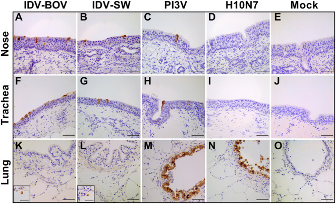
FIGURE 3.

Immunohistochemical analysis of infected explants. Each image represents nasal (A–E), tracheal (F–L), or lung (K–O) ovine explants infected with IDV isolated from bovine (IDV-BOV) or swine (IDV-SW), parainfluenza 3 virus (PI3V), avian influenza H10N7 (H10) or mock infected and collected at 72 h p.i. In the images (M,N), the insertion shows a magnification of a positive alveolar macrophage in lung explant, respectively, infected by IDV-BOV and IDV-SW. Scale bar = 50 μm.
