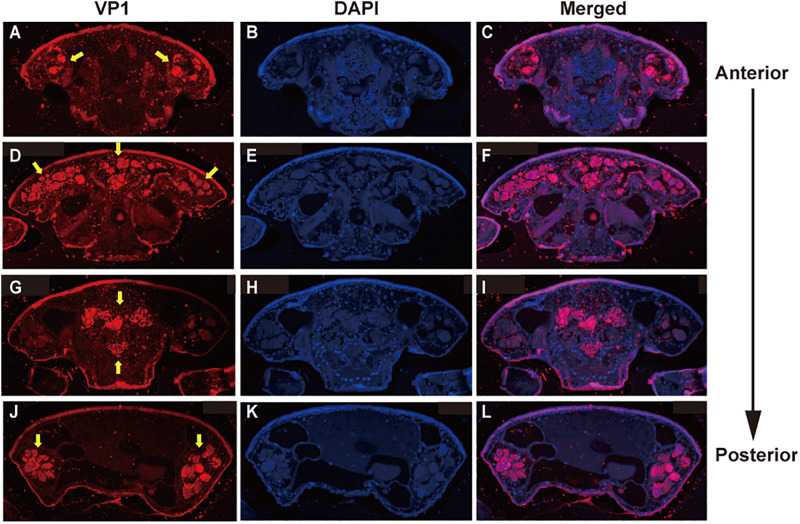
FIGURE 3.
Localization of DWV in T. mercedesae. Transverse sections of T. mercedesae were immunostained by anti-VP1 antibody (VP1: A,D,G,J) as well as DAPI (B,E,H,K). The merged images (Merged: C,F,I,L) are also shown. VP1-positive large dense spheres are indicated by yellow arrows. The dorsal side of mite is up and the anterior to posterior direction of mite body is also shown.

