
Figure 1.
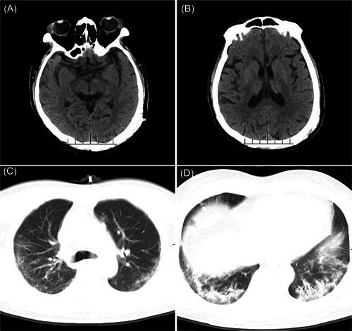
Head CT and chest CT of the patient. A,B, No obvious abnormalities were found on brain CT. C,D, Diminished bilateral lung translucence, with multiple ground‐glass opacities distributed in both in the middle and lateral fields of the lung. Multiple cord‐like shadows can be observed in the bilateral lower lungs, with interlobular septal thickening and bronchiectasis. CT, computerized tomography