
Fig 2.
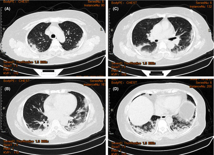
Typical CT images of patients with severe 2019 novel coronavirus pneumonia (COVID‐19). Strip‐like consolidation shadows and patchy ground glass‐like density shadows can be seen in the apical and posterior segment of the upper lobe of both lungs, tongue segment of the left upper lobe, and the lower lobe of both lungs with a fuzzy boundary, mainly distributed under the pleura of both lungs when the patient was in severe condition. [Colour figure can be viewed at http://www.wileyonlinelibrary.com/]