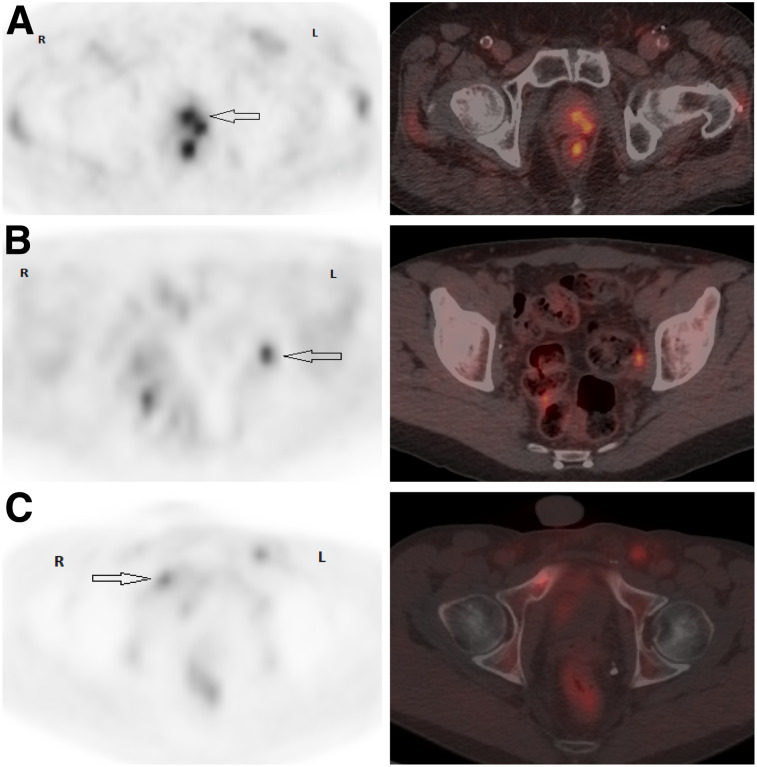
FIGURE 4.
Patient examples of pelvic axial PET and PET/CT images. (A) PCa 4.3 y after intensity-modulated radiotherapy (Gleason 7 [4 + 3]; T2cN0M0; PSA, 2.28 ng/mL). PET/CT shows suspected left posterior prostatic uptake (SUVmax, 3.3), scored 2 (arrow). Local biopsy was positive for PCa. (B) PCa 6.4 y after RP plus pelvic lymph node dissection (Gleason 8 [4 + 4]; pT2bN0M0; PSA, 0.48 ng/mL). PET/CT shows suspected focal uptake (SUVmax, 4.5) in nonenlarged left obturator lymph node, scored 2 (arrow). Histology was positive for PCa. (C) PCa 10 mo after RP plus pelvic lymph node dissection (Gleason 8 [4 + 4]; pT4N1M0; PSA, 0.46 ng/mL; PSA doubling time, 2.1 mo). PET/CT shows 2 suspected bone foci, one in right pubic ramus (SUVmax, 3) with sclerotic lesion on CT (arrow) and the other in posterior eight rib (not shown), scored 2. Biopsy of right pubic ramus was positive for PCa.

