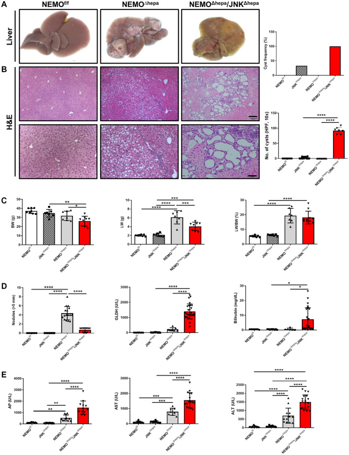
Fig. 1.

Deletion of Jnk1/2 in 52‐week‐old NEMOΔhepa livers triggers cyst formation. (A) Macroscopic view of livers from 52‐week‐old NEMOf/f (wild type), NEMOΔhepa, and NEMOΔhepa/JNKΔhepa mice. (B) Representative H&E staining of liver sections of NEMOf/f, NEMOΔhepa, and NEMOΔhepa/JNKΔhepa livers at 52 weeks of age. Different magnifications were used (left). Scale bar 200 µm. Cyst frequency and number of visible microscopic cysts per 10× view field were calculated and graphed (right), magnification is 10× for upper and magnification is 20× for lower. (C) BW (left); LW (center); LW/BW ratio (right). (D) Tumor burden for each individual mouse was characterized by calculating total number of visible tumors >5 mm in diameter per mouse (left); serum levels of GLDH (center); bilirubin (right). (E) Serum levels of AP (left), AST (center), and ALT (right), in 52‐week‐old NEMOf/f, JNKΔhepa, NEMOΔhepa, and NEMOΔhepa/JNKΔhepa mice. Data are presented as mean ± SEM. *P < 0.05; **P < 0.01; ***P < 0.001; ****P < 0.0001. Abbreviations: AP, alkaline phosphatase; GLDH, glutamate dehydrogenase; H&E, hematoxylin and eosin.
