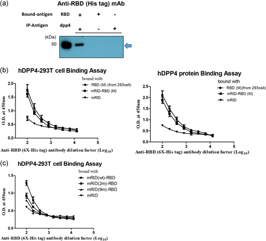
Figure 3.

Binding between RBD and hDPP4. (a) Co‐immunoprecipitation analysis of MERS RBD (2 μg, Escherichia coli) binding to hDPP4 protein (2 μg, 293 cell) using protein G (50 μg/μl) and hDPP4 specific antibody. RBD protein (his tagged) was detected by western blot analysis using anti‐6 × ‐His tag monoclonal antibody. (b) Dose‐dependent binding of His‐tagged RBD (5 μg/ml) from E. coli and 293T cell (eEnzyme), respectively, with hDPP4 (recombinant or ectopically displayed in 293T cells) as coating antigen determined by ELISA. RBD(M) (derived from E. coli) was compared with mRID (the negative control) and RBD from 293T cells (the positive control). Three independent experiments were performed (n = 3). (c) Binding ELISA of mRID (wild‐type (wt), 2 m, 9 m)‐RBD and 293T cells overexpressing hDPP4. The ELISA data were obtained in duplicate. The antibodies were twofold serially diluted from 1:100. All data are presented as mean ± SD. ELISA, enzyme‐linked immunosorbent assay; K, Korean strain; M, Middle East strain; MERS, Middle East respiratory syndrome; mRID, mouse RNA‐interacting domain; RBD, receptor‐binding domain [Color figure can be viewed at wileyonlinelibrary.com]
