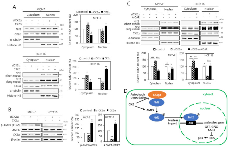
Fig. 4.
CK2 downregulation reduces the nuclear localization of Nrf2 through inhibiting AMPK in human cancer cells. (A) Cytoplasmic and nuclear fractions were isolated from the cells transfected with CK2α siRNA or pcDNA3.1-HA-CK2α and then both extracts were analyzed by immunoblotting (left panels). α-Tubulin (cytoplasmic marker) and histone H3 (nuclear marker) levels were quantified as loading controls. Graphs present the quantification of Nrf2 levels relative to the subcellular marker levels (right panels). (B) Cells were transfected with CK2α siRNA or pcDNA3.1-HA-CK2α for 48 h, lysed, and electrophoresed on a 10% SDS–polyacrylamide gel. Protein bands were visualized by immunoblotting (upper panel). Graphs show the quantification of the p-AMPK levels relative to AMPK level (bottom panel). (C) Cytoplasmic and nuclear extracts were isolated from the cells, and then both extracts were analyzed by immunoblotting (upper panels). The levels of α-tubulin (cytoplasmic marker) and histone H3 (nuclear marker) were quantified as loading controls. Graphs represent the quantification of Nrf2 levels relative to subcellular marker levels (bottom panels). All the data are shown as means ± SEM. *P < 0.05; **P < 0.01; ***P < 0.001. (D) Mechanistic model illustrating the involvement of Nrf2 in the CK2 downregulation-induced cellular senescence.

