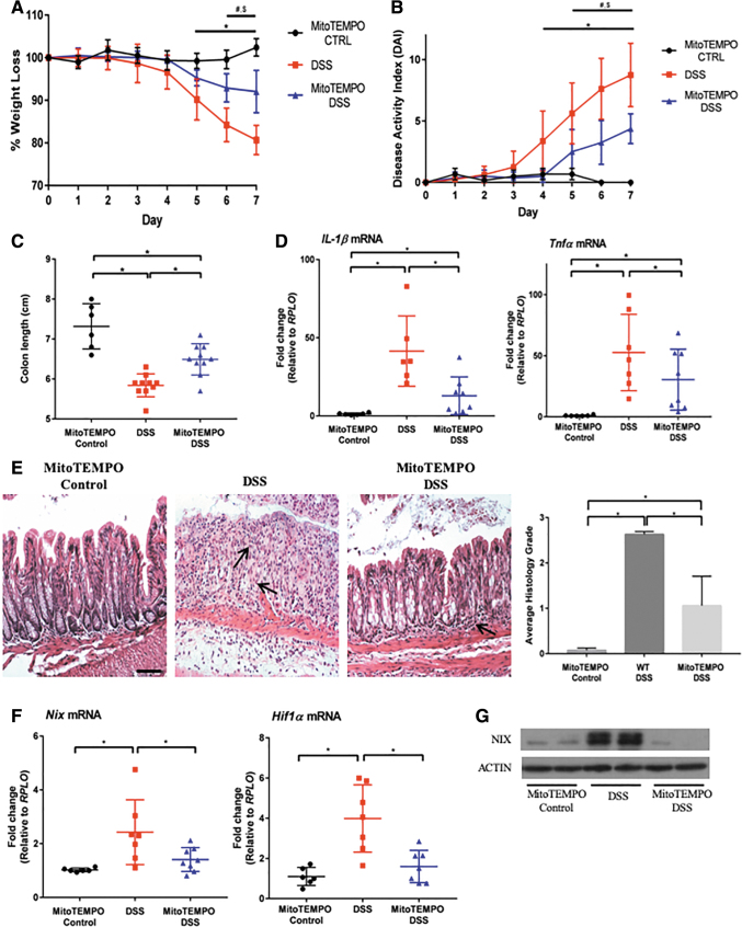
FIG. 6.
MitoTEMPO protects from DSS-induced colitis by scavenging mitochondrial ROS in vivo. (A) Mice were subjected to 3% DSS for 7 days, with MitoTEMPO administered daily via intraperitoneal injection. MitoTEMPO-treated mice were protected from weight loss and exhibited a lower DAI score (B) than DSS only. Significant changes were observed between MitoTEMPO control versus 3% DSS mice (*p ≤ 0.05), MitoTEMPO control versus MitoTEMPO 3% DSS (#p ≤ 0.05), and 3% DSS versus MitoTEMPO 3% DSS ($p ≤ 0.05) (n = 6 control, n = 10 DSS, n = 10 MitoTEMPO DSS). (C) MitoTEMPO 3% DSS mice also displayed longer colons than DSS only mice (n = 6 control, n = 10 DSS, n = 10 MitoTEMPO DSS). (D) Inflammatory cytokines were analyzed via qPCR, showing a reduction in Il-1β and Tnfα in MitoTEMPO 3% DSS mice (n = 6 control, n = 7 DSS, n = 8 MitoTEMPO DSS). (E) H&E staining shows a preservation of structure and a decrease in inflammatory infiltrate in MitoTEMPO 3% DSS mice with a significant decrease in the histological scoring, as performed by a blinded pathologist (20 × magnification, scale bar = 100 μm) (n = 6 control, n = 10 DSS, n = 10 MitoTEMPO DSS). Arrows point out areas of immune cell infiltration. (F) Nix and Hif1α were significantly decreased in MitoTEMPO 3% DSS-treated mice when compared with DSS only mice (n = 6 control, n = 7 DSS, n = 8 MitoTEMPO DSS). (G) Protein lysates were analyzed via Western blot, showing a decrease in NIX in MitoTEMPO 3% DSS-treated mice compared with DSS alone (n = 2 for all groups). Blots are representative of several experiments. Please note that the y-axis in (A) and (C) are not set to 0 to aid in the visual representation of the data. *p ≤ 0.05. H&E, hematoxylin and eosin.

