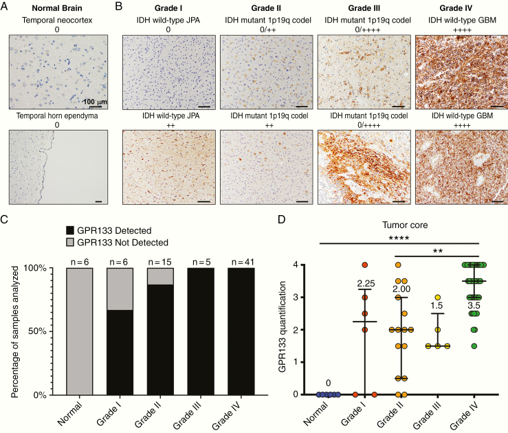
Figure 2.
Non-neoplastic brain tissue lacks GPR133 expression, whereas gliomas express GPR133 at higher levels with increasing WHO grade. (A) GPR133 immunohistochemistry in temporal neocortex and temporal horn ependyma in representative control specimens. Scale bar, 100 μm. (B) GPR133 antibody stains in gliomas grade I–IV. Scale bar, 100 μm. (C) Proportion of samples with GPR133 expression by WHO grade. (D) Level of GPR133 expression by grade (P < .0001, Kruskal–Wallis test; post hoc Dunn’s **P < .003; ****P < .0001). codel: codeleted.

