Figure 3.
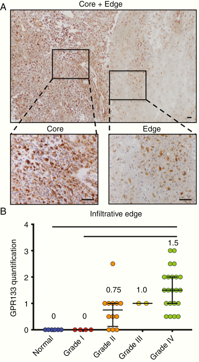
GPR133 is detected in both the core and infiltrative edge of gliomas, as well as in areas of pseudopalisading necrosis. (A) GPR133 immunohistochemistry in IDH wild-type GBM tissue at the core (left zoom) and infiltrative edge (right zoom). The tumor core was graded as +++, and the tumor edge was graded +/++. Scale bar, 100 μm. (B) GPR133 quantification at the infiltrative edge by grade (P < .0001, Kruskal–Wallis test; post hoc Dunn’s *P < .01; **P < .001). Scale bar, 100 μm.

