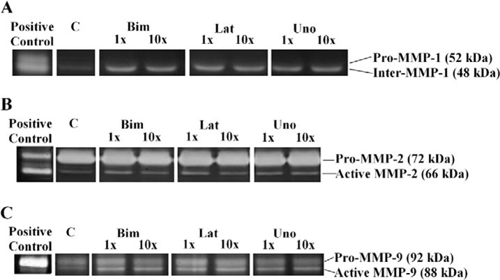
Figure 3.
Representative zymography for pro- and active forms of MMPs-1, −2, and −9 in human TM endothelial cells incubated with free acid moieties bimatoprost, latanoprost, and unoprostone for 24 hours. Active form of MMP-3 was not detected. C represents control, 1x and 10x represent samples incubated at the peak aqueous concentrations and ten times the peak aqueous concentration of these drugs, respectively, for 24 hours.

