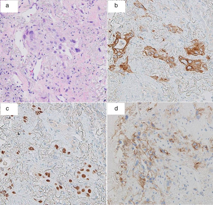
Figure 3.

Pathological findings of tumor tissue obtained by CT‐guided needle biopsy showed squamous cell carcinoma. (a) Hemotoxylin‐eosin stain revealed that the right lung mass consisted of atypical squamous cells, which was partially positive for (b) cytokeratin 5/6 and (c) p40. (d) Furthermore, programmed death‐ligand 1 (PD‐L1) showed high expression with a tumor proportion score (TPS) >75%.
